When “Charcot Foot” Means Two Very Different Things
by Kenneth Raymond and John Rahman, C.O. (C) F.C.B.C.
There is a name in medicine that appears in more places than almost any other. It sits at the top of a peripheral neuropathy that affects more than 3 million worldwide. It describes a joint disease that destroys bone and architecture in the insensate foot. It marks a pair of clinical triads, one for multiple sclerosis and one for cholangitis. It names a sign of intermittent claudication: a crystal found in the sputum of asthma patients (Charcot-Leyden Crystals). The name is attributed to amyotrophic lateral sclerosis (ALS) in Europe for well over a century. It belongs to an island in Antarctica.
The name is Charcot, and the man behind it, Jean-Martin Charcot (1825-1893), has been described by more than one historian of medicine as the most eponymously productive clinician in the history of the field (Goetz, 1987; Waraich & Shah, 2018)13, 25. There is more in medicine attributed to Charcot than perhaps any other individual.
Charcot’s name is formally associated with at least 15 medical eponyms, a count acknowledged by many peer-reviewed papers in the academic literature (Goetz, 1987; Waraich & Shah, 2018)13, 25. Known in French medical history as le père de la neurologie, the father of neurology, and dubbed by contemporaries “the Napoleon of the Neuroses,” Charcot built his legacy at the Hôpital de la Salpêtrière in Paris, where he oversaw a population of roughly 5,000 patients, nearly 3,000 of whom carried a neurological diagnosis (Teive et al., 2011)23.
Charcot did not merely describe diseases. He catalogued them, classified them, illustrated them, and taught them in theatrical public demonstrations that drew the physicians and intellectuals of Paris, and those traveling from across Europe, to his amphitheatre. Among the students who came to study under him were Sigmund Freud, Joseph Babinski, and Georges Gilles de la Tourette, whose own eponymous syndrome Charcot himself named (Waraich & Shah, 2018)25.
All of this is relevant because Charcot’s name also generates confusion in the Charcot-Marie-Tooth disease (CMT) community. This confusion centers on two presentations of the foot. One belongs to CMT, and one does not, except in rare circumstances. These two are not the same thing and do not share any mechanism. They do not look alike, progress alike, or require the same care. The name? Charcot foot.
Charcot foot, also known medically as Charcot neuroarthropathy (noo-ro-ar-THROP-ah-thee) and, in the literature, as Charcot neuropathic osteoarthropathy or simply CN, frequently surfaces in community conversations, social media posts, and sometimes in clinical settings, often interchanging with the foot deformities CMT often causes. These two presentations are distinctly distinct, and confusing the two is not without consequence. Caring for either is often patient-initiated through self-advocacy, and treatment outcomes depend on knowing the distinctions.
One Name, Two Feet
CMT is an inheritable peripheral neuropathy, meaning a disease of the peripheral nervous system. The peripheral nervous system (PNS) is the vast network of nerves that lie outside of the brain and spinal cord. CMT is also a neuromuscular disease because damage to the peripheral nerves results in consequences in the muscles they control.
CMT is a disease of enormous genetic complexity, caused by mutations in more than 140 genes identified to date, which give rise to more than 170 genetically distinct subtypes (Experts in CMT, 2026)11. Despite this heterogeneity, one feature is remarkably consistent across the CMT spectrum: the feet are affected, often in a recognizable way.
CMT’s recognizable foot presentation, informally the “CMT foot,” is a product of nerve dysfunction and is built by muscle imbalance. It is a structural consequence of the disease, and it has a clinical name: cavovarus (kav-oh-VAIR-us) or cavoequinovarus (kav-oh-eh-KWEE-no-VAIR-us) foot deformity, which is more commonly called pes cavovarus. Charcot foot is something categorically different.
Charcot foot is a destructive disease of the bones and joints of the foot and ankle driven primarily by sensory neuropathy and the loss of protective pain sensation, culminating in progressive bone fragmentation, joint collapse, and severe deformity that, in its worst presentation, can lead to amputation (Rogers et al., 2011; Trieb, 2016)21, 24. It can occur in CMT, but it is rare. And it occurs commonly in many conditions that have nothing to do with CMT, most prominently in diabetes mellitus.
The two presentations bear the Charcot name for the same reason they carry the same man’s legacy in different medical domains: Jean-Martin Charcot described both. He described the inherited neuropathy that bears his name alongside Pierre Marie (Charcot’s apprentice) and Howard Tooth in 1886 (Charcot & Marie, 1886)4. He described neuroarthropathy, the joint destruction driven by sensory neuropathy, eighteen years earlier in 1868, in patients with tabes dorsalis, the neurological manifestation of tertiary syphilis (Charcot, 1868)3. One name. Two diseases. An entirely understandable source of confusion, particularly when the CMT community is already navigating the complexity of Charcot’s name appearing in the name of their own disease. This confusion has a geographic dimension worth acknowledging.
Charcot’s Influence on Medicine
In France and in several parts of continental Europe, Charcot’s influence on medicine is so deeply embedded in clinical culture that his name is used more freely and casually as medical shorthand than in North America. ALS is routinely called la maladie de Charcot in France, as it has been since Charcot first described and named it in 1874 (Teive et al., 2011)23. In this context, some European clinical and patient literature uses “Charcot foot” as a broader shorthand for neuropathic foot pathology, including, at times, the “CMT foot” cavovarus presentation. This is not technically incorrect from a historical standpoint since Charcot described both. But it has contributed significantly to the confusion that circulates globally in the CMT community, where the question “do I have Charcot foot?” sometimes means “do I have the bone-destroying joint disease?” and sometimes means “do I have the high-arched, twisted, crooked, structurally deformed foot that CMT causes?” The answer to these questions requires the dismantling of what each presentation actually is, how each is built, and what each looks like.
The CMT Foot: Built From the Inside Out
Charcot neuroarthropathy is not a deformity beset by muscle imbalance. It is a deformity of destruction.
To understand the CMT foot, it helps to understand what CMT is doing at the nerve and muscle levels. CMT attacks the peripheral nerves, the long communication lines that carry instructions from the spinal cord out to the muscles and carry sensory information back from the skin and joints to the spinal cord and brain. In the most common CMT subtypes, this damage is demyelinating: the myelin (MY-eh-lin) sheath, the insulating layer that wraps around nerve fibers and allows signals to travel quickly and reliably, is compromised (Dyck et al., 1963)10. In axonal subtypes, it is the nerve fiber itself, the axon (AX-on), that degenerates.
In either case, what typically takes place is that the muscles connected to the furthest ends (distal) of the longest nerves are more severely impacted by CMT’s effects. And over time, these effects progress to what’s connected to shorter nerves, closer to the spinal cord (proximal).
The muscles of the foot and lower leg are served by these long peripheral nerves, and when the nerves degrade, the muscles weaken. But they do not all weaken at the same rate or at the same time. This selective, asymmetric weakening is the engine that builds the CMT foot. If we look at the foot as a system of opposing forces, we can get inside of what causes CMT’s foot deformities.
Unequal Pull Equals Unequal Balance Equals CMT Foot
Think of the foot’s architecture as a tent held up by ropes pulling in multiple directions. As long as all the ropes maintain roughly equal tension, the tent holds its shape. The moment some ropes go slack while others remain taut, the tent distorts in the direction of the surviving tension. This is exactly what happens in CMT.
In CMT, the muscles of the anterior and lateral compartments of the leg, specifically the tibialis anterior (tib-ee-AH-lis an-TEER-ee-or) and the peroneus brevis (peh-ROH-nee-us BREV-is), are among the first to weaken (Mann, 1992; Krähenbühl & Weinberg, 2019)15, 14. These muscles normally act as important counterbalances. The tibialis anterior lifts the front of the foot upward during walking, preventing the front of the foot from dropping (foot drop), and also helps resist the downward pull on the inner arch. The peroneus brevis, running along the outside of the ankle, pulls the foot outward into eversion (outward and away from the body’s midline), and counterbalances the foot’s tendency to roll inward.
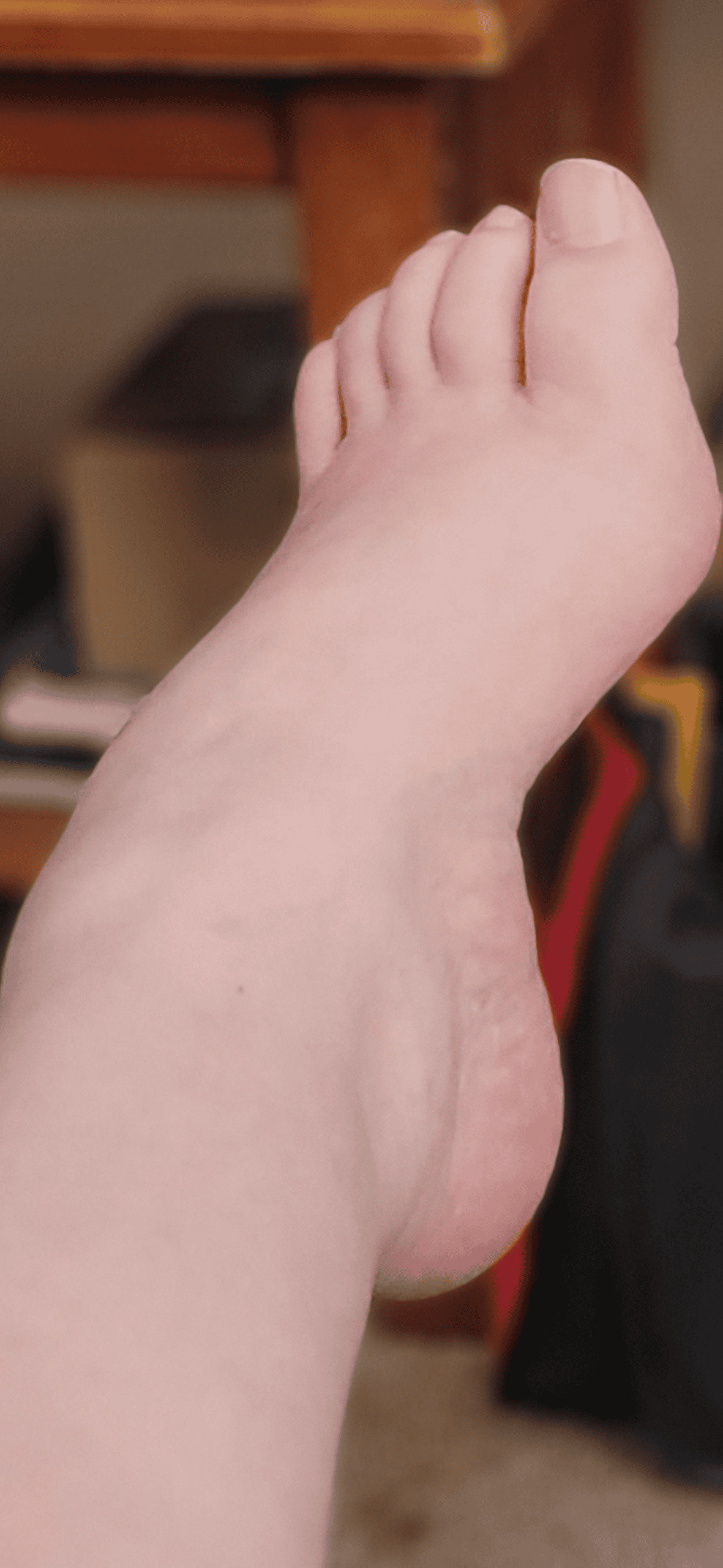
When these two muscles weaken in CMT, their opposing muscles, the peroneus longus (peh-ROH-nee-us LON-gus) and the tibialis posterior (tib-ee-AH-lis pos-TEER-ee-or), continue to function at their original strength, at least initially (Krähenbühl & Weinberg, 2019; Acquired Pes Cavus in CMT, 2016)14, 1. The peroneus longus, now unopposed by the weakened tibialis anterior, pulls the first metatarsal (meh-tah-TAR-sal), the long bone at the base of the big toe, downward toward the ground. This plantarflexes (plan-tar-FLEX-es, meaning bends downward) the first ray, which is the column of bones running through the big toe. The forefoot tilts inward. The inner arch, already under the pull of an unbalanced peroneus longus and the contracting plantar fascia (PLAN-tar FAY-shee-ah, the thick band of connective tissue spanning the sole), begins to rise. At the same time, the tibialis posterior, no longer balanced by the weakened peroneus brevis, pulls the foot into inversion, rolling it inward toward the midline (Mann, 1992)15.
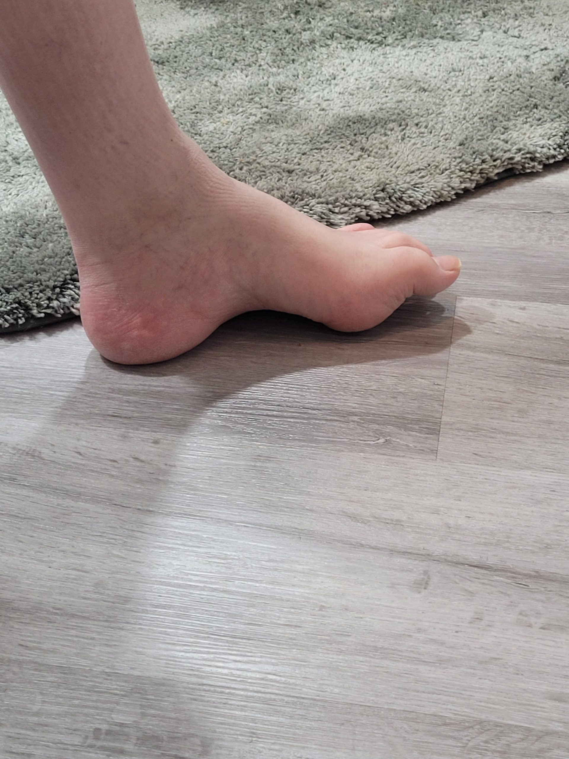
The tripod of the foot, meaning the heel, the ball of the foot under the big toe, and the ball of the foot under the little toe, loses its flat, even contact with the ground. The front inside edge drops, the rear of the foot rolls inward, and the arch climbs. The intrinsic muscles of the foot, which are the small muscles that control toe position, develop contractures, and the long toe extensors, recruited to assist with ankle lift when the tibialis anterior has failed, begin to pull the toes upward into a claw-toe deformity (hammertoes) (Pes Cavus StatPearls, 2023)20. What emerges is the cavovarus foot: a high, rigid arch (cavus), combined with the heel turned inward under the ankle (varus).
When equinus is also present, the tendency of the foot to point downward at the ankle, driven by a tight Achilles tendon, results in a pes cavovarus presentation. The foot seems perpetually trying to walk on its outer edge. The heel strikes the ground on its outer side. Lateral ankle instability is common, as the outer ankle ligaments are chronically overloaded (Pes Cavus ACNR, n.d.)19. Stress fractures of the fifth metatarsal are a recognized complication of this lateral overloading. This is the CMT foot.
The CMT foot is a bilateral deformity (bilateral = both right and left), develops slowly, and progresses gradually over years and decades (Pes Cavus StatPearls, 2023)20. It is shaped by motor nerve damage and the muscle imbalance CMT creates. The bones themselves undergo structural changes (bone morphology), especially in the calcaneus (heel bone). Sensory loss does contribute to CMT in many individuals, particularly in later disease stages, but the cavovarus deformity itself is primarily a motor phenomenon. It is built by muscles pulling in the wrong directions, not by sensory loss removing the body’s ability to protect its joints. Stress fractures can readily occur from these unbalanced forces enacted on the foot by CMT, but there is no bone fragmentation or structural collapse of the joint spaces. This is a key distinction when considering Charcot foot.
Charcot Neuroarthropathy: Built From the Outside In
To dismantle Charcot foot, start by examining what pain actually does to the body during movement. Every step you take places mechanical stress on and through the bones and joints of the foot. Normally, the nervous system monitors this stress through pain receptors and proprioceptors (pro-pree-oh-SEP-tors, specialized nerve endings that detect position, pressure, and damage). When stress approaches or exceeds safe limits, the body responds by shifting weight, adjusting gait, stopping walking, or sending a sharp pain signal that demands attention. This feedback loop is the body’s first line of protection for its own skeletal architecture.
The feedback loop operates constantly and mostly unconsciously. It’s what causes you to wince when you step on a pebble with a bare foot, and it is what prevents the cumulative mechanical damage of daily mobility from destroying the bones and joints of a normal foot. In Charcot Neuroarthropathy (CN), however, this protective system is severely compromised or even absent.
The fundamental requirement for CN is peripheral neuropathy severe enough to eliminate the protective pain sensation and proprioception feedback loop in the affected limb (Trieb, 2016; Rogers et al., 2011)24, 21. Without this feedback loop, the foot sustains repetitive microtrauma during ordinary activities, such as walking, standing, and going about daily life, without the person feeling it, without the body responding to it, and without it stopping. The damage accumulates silently.
Two theories have long been proposed to explain the pathophysiology of CN (its underlying biology). The neurotraumatic theory (noo-ro-TRAW-mat-ik) holds that the loss of sensory feedback allows repeated mechanical injury to accumulate unchecked (Charcot Neuropathic Osteoarthropathy StatPearls, 2024; Charcot Arthropathy Medscape, n.d.)7, 5. Microtrauma that a healthy foot would detect, respond to, and recover from instead compounds over time, leading to progressive bone fracture and joint instability. The neurovascular theory (noo-ro-VAS-kyu-lar) proposes a second mechanism
The neurovascular theory of CN proposes that autonomic neuropathy disrupts sympathetic control of blood flow to the foot, leading to hyperemia (hy-per-EE-mee-ah, an abnormally elevated blood flow), which activates osteoclasts (OS-tee-oh-CLASTS, the cells that break down and resorb bone tissue) (Dardari, 2020)9. The current understanding is that both mechanisms likely contribute, with contemporary research emphasizing the role of the RANK/RANK-L signaling pathway, a molecular cascade that, when dysregulated by local inflammation, drives excessive osteoclastic activity and accelerated bone resorption (Charcot Neuropathic Arthropathy PMC, 2016; Montechi et al., 2021)6, 17. The result, in whatever combination of mechanisms produces it, is a foot undergoing active bone destruction.
In the acute phase of CN, the foot becomes swollen, warm, and red, often dramatically so. The heat is measurable. An infrared thermometer held over an acutely active Charcot foot will commonly show a temperature difference of 2 degrees Celsius or more compared to the unaffected side (Rogers et al., 2011)21. The presentation is so inflammatory that it is frequently misdiagnosed as cellulitis, a misdiagnosis that delays appropriate off-loading intervention and accelerates joint destruction (Charcot Neuroarthropathy in Acute Setting PMC, 2023)8.
Pain, counterintuitively, may be present in CN despite the sensory neuropathy. Many patients report a dull, deep ache during weight bearing that does not fully capture the extent of damage beneath the skin, though others report little pain relative to the severity of what imaging reveals (Rogers et al., 2011)21.
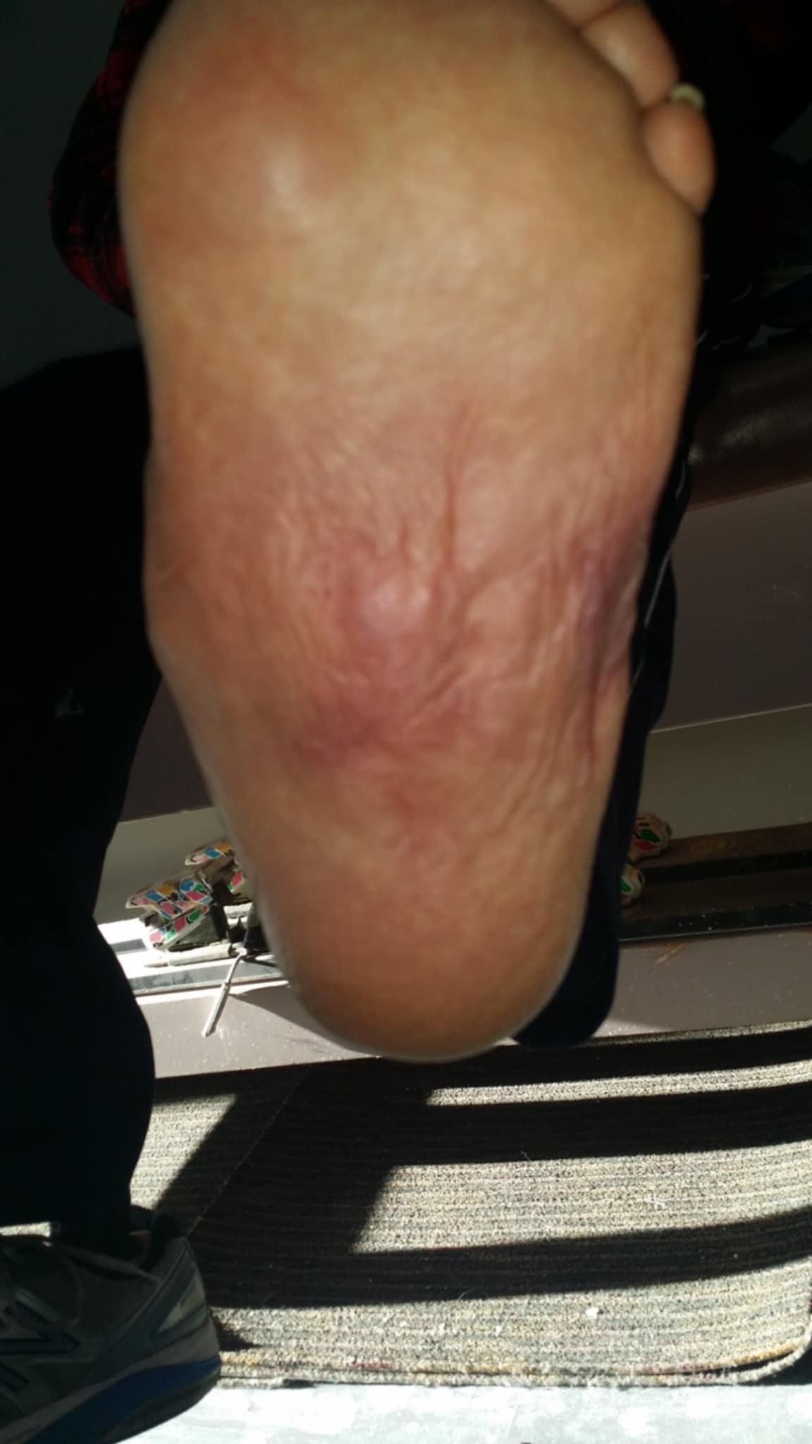
As CN progresses through its stages, the bones of the midfoot, most commonly the tarsometatarsal (TAR-so-meh-tah-TAR-sal) joints, which account for roughly 60% of CN presentations, fragment, displace, and collapse (Rogers et al., 2011)21. The arch of the foot, rather than rising as in the CMT cavovarus presentation, collapses under the weight of the body, pressing through bones that can no longer support it. In the most classic and severe presentation, the midfoot collapses completely downward, producing a “rocker-bottom” deformity.
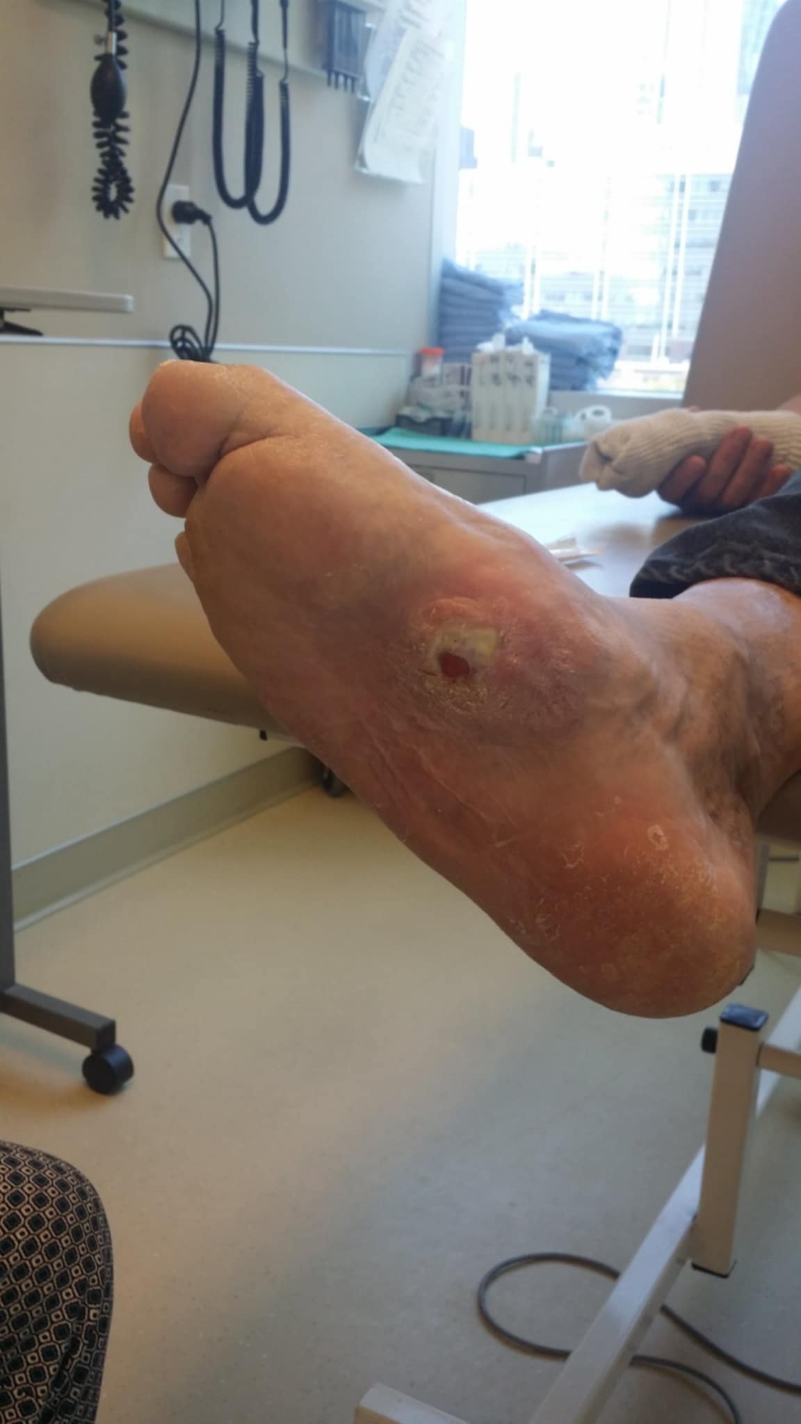
The name “rocker-bottom” comes from the collapsed midfoot bowing downward and creating a curved sole that rocks like the base of a rocking chair. A bony prominence forms at the bottom of the collapsed midfoot, and that prominence, constantly in contact with footwear and the ground, is at severe risk for pressure ulceration (open wound). Pressure ulcers in an insensate (doesn’t transmit pain signals) foot can quickly become infected, progressing to osteomyelitis (os-tee-oh-my-eh-LY-tis, a severe bone infection) and, in severe cases, requiring amputation (Charcot Foot in Diabetes, Diabetes Care, 2011)21. This is fundamentally different from the CMT foot.
The CMT foot typically has a high arch. The Charcot foot, in its classic midfoot presentation, has a collapsed arch or no arch. Individuals with CMT can also have flat feet (pes planus), but this is still due to muscle imbalances, unlike CN.
The CMT foot is built over years by muscle imbalances that can change the shape of the bones, but they remain structurally intact. The Charcot foot, in contrast, is built by the destruction of bone architecture in the absence of protective sensation. One is a problem of shape. The other is a survival problem.
Charcot Neuroarthropathy in CMT: The Rare Intersection
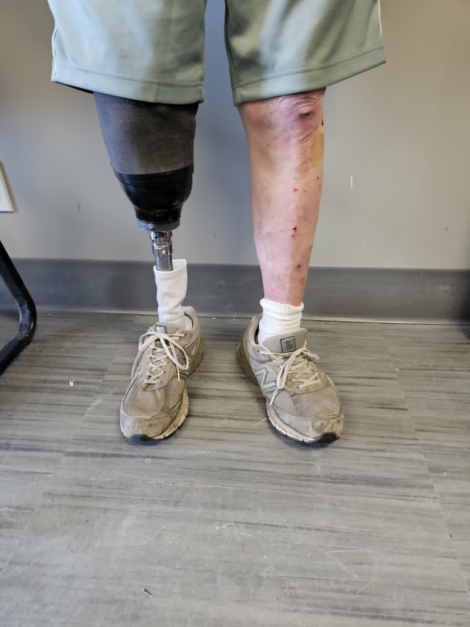
Charcot neuroarthropathy can occur in CMT. The published case literature, though sparse, confirms this. Singh et al. (2021)22, in a case series published in Foot and Ankle Surgery, described three patients with CMT1A who developed CN, including one who underwent a transtibial (below-the-knee) amputation as a consequence of bilateral midfoot CN. The paper is notable in that it involved co-authors from both the Royal National Orthopaedic Hospital and the UCL Queen Square Institute of Neurology’s MRC Centre for Neuromuscular Diseases, lending it significant clinical authority relevant to CMT.
Parks and Benstead (2010)18 described a case of Charcot ankle arthropathy in a CMT1A patient in whom type 2 diabetes mellitus was present and explicitly described it as exacerbating the presentation. Earlier case reports, including those by Bruckner and Kendall (1969)2 and Medhat and Krantz (1988)16, documented CN development in CMT patients, with the latter describing CN emerging at the ankle following triple arthrodesis to correct CMT foot deformities.
The existing literature collectively characterizes CN in CMT as rare. The published case count in the peer-reviewed literature remains countable in the single digits, even after more than 50 years of occasional documentation. Singh et al. (2021)22 explicitly note that patients with CMT are at risk for developing Charcot joints, and that this risk exists even in the more common CMT1A, where sensory deficits, though present on clinical examination, may not be subjectively prominent to the patient. This is an important point.
CMT is a disease of the peripheral nervous system that, depending on subtype and individual presentation, can affect motor nerves, sensory nerves, or both (sensorimotor polyneuropathy). Pain sensation and impaired proprioception are well-documented and progress over time (Gemignani et al., 2004)12. It is this sensory component, when sufficiently severe, that creates the physiological conditions under which CN can develop.
Why CN in CMT is rare, while it is common in conditions like diabetes, likely reflects the nature and severity of the sensory deficit involved. Diabetes produces a predominantly sensory peripheral neuropathy that, over years of poor glycemic control, can eliminate protective sensation feedback loops profoundly and comprehensively. CMT, even in subtypes with significant sensory involvement, tends to preserve some degree of sensory feedback for longer, and its autonomic involvement, the other component implicated in CN’s neurovascular mechanism, is less prominent than in diabetic neuropathy (Singh et al., 2021)22. Nevertheless, rare does not mean impossible. Any patient with CMT who has a hot, swollen foot should not have CN summarily dismissed without appropriate clinical evaluation.
What Each Foot Looks Like: The Structural and Visual Distinction
The differences in mechanism help explain the differences in appearance, but it is worth being explicit, because these two presentations do not look alike.
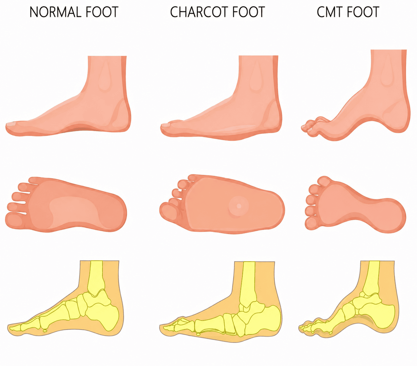
The CMT foot, cavovarus or pes cavovarus, is characterized by a high, rigid, elevated arch on the medial (inner) side of the foot. The heel is turned inward under the ankle, pointing toward the midline of the body rather than straight down. The toes are often clawed (hammertoes), with the joints hyperextended at the metatarsophalangeal level (base of the toes) and flexed at the interphalangeal level (the toe knuckles). The forefoot is tilted inward, and the outside edge of the foot is often the primary weight-bearing surface during ambulation (a “supinated” position). The arch is not only high but also rigid. It does not flex appreciably with weight-bearing (the arch of an unaffected, normal foot functions as a shock absorber, flexing under weight loading). Calluses form under the metatarsal heads, particularly the first and fifth, and under the ball of the foot, reflecting the abnormal pressure distribution.
The CMT foot deformity is usually bilateral and relatively symmetric, though asymmetry in severity is common. The foot’s bones are intact, albeit misshapen. Imaging does not show fragmentation. The problem is configuration and shape, not architecture. CN, in its most common midfoot presentation, looks dramatically different.
The Charcot foot arch does not rise. It collapses or disappears. In advanced CN, the foot widens, and the midfoot may bulge downward, creating the rocker-bottom profile described above. The foot becomes visibly broader than normal. In the acute phase (beginning, short-term), the entire foot or a region of it is swollen, warm to the touch, and may appear red or dusky. In later stages, the warmth resolves as the acute inflammatory phase ends, but the deformity remains and may be severe.
Fractured and displaced tarsal and metatarsal bones, dislocated joints, and collapsed bony columns worsen as CN progresses. Imaging reveals the internal destruction through chronic (long-term) fracture lines, bone fragmentation, joint subluxation or dislocation, and, in chronic cases, sclerosis (hardening) and remodeling of destroyed bone. The bones themselves have failed. The problem is not a configuration issue but a catastrophic structural compromise.
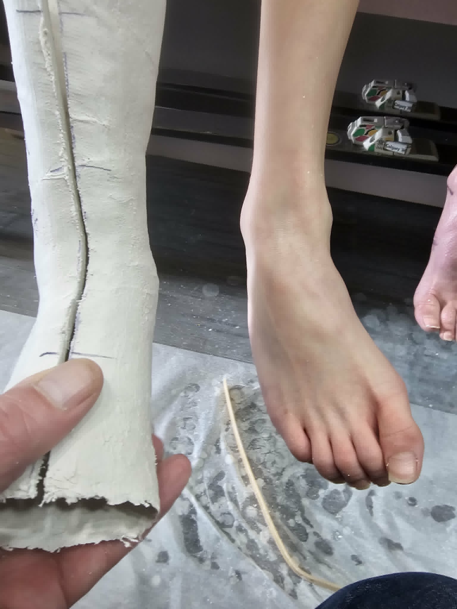
“When I’m fitting a patient with the CMT foot, I’m managing a structural deformity that has altered the foot’s mechanics,” said John Rahman, C.O. (c) F.C.B.C., founder Rahman Orthotics in Calgary, Alberta. “I’m accommodating an arch that’s too high, redistributing pressure away from overloaded areas, and controlling the inward rotation of the hindfoot. When I’m managing a Charcot foot, I’m dealing with a foot whose load-bearing architecture has been partially or wholly destroyed. The goals are entirely different.”
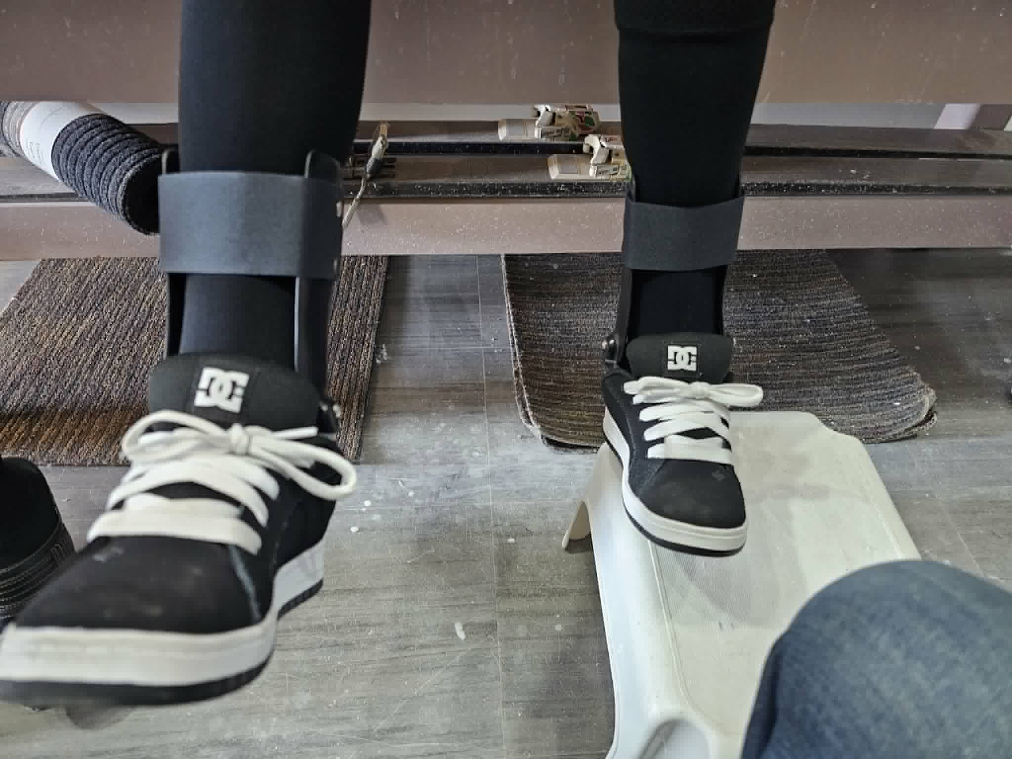
John continues, “The CMT foot needs mechanical correction and support. The Charcot foot, especially in its acute phase, needs off-loading, protection from further destruction, and close monitoring for ulceration. Confusing the two is not just a semantic error. It can lead to the wrong care, and in the case of an active Charcot process, to irreversible harm.” This clinical difference in management is why the distinction matters far beyond terminology.
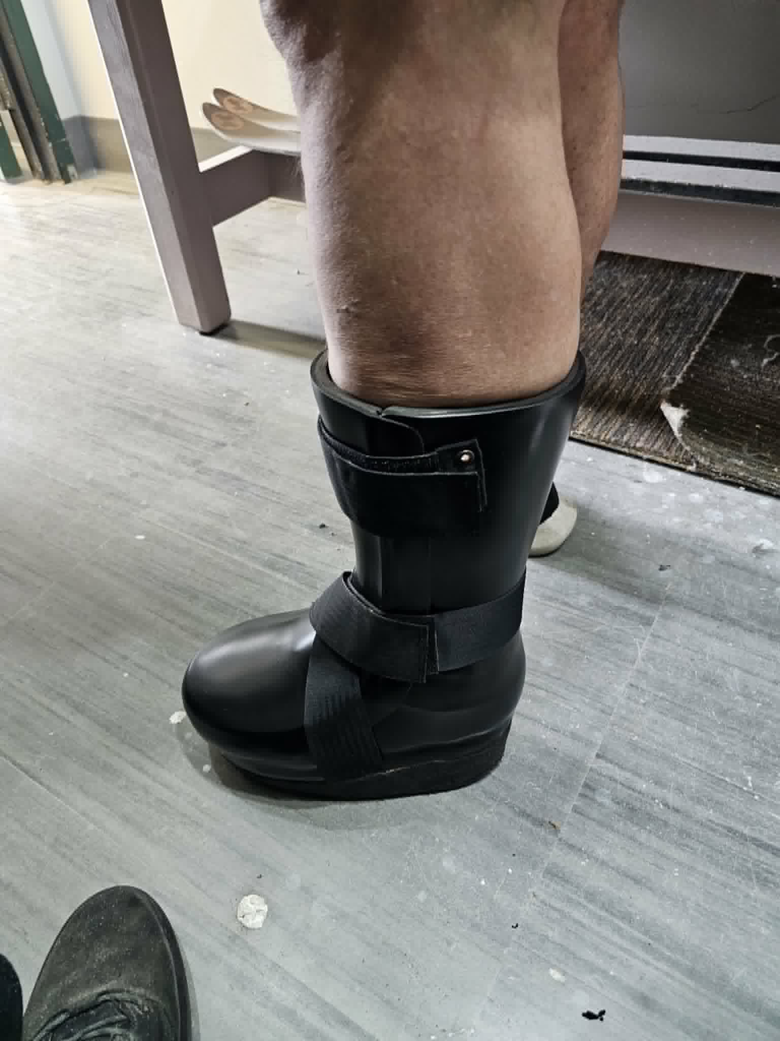
The active Charcot foot requires urgent off-loading, typically total contact casting or equivalent non-weight-bearing immobilization, to stop the destructive cascade and allow the inflammatory phase to resolve (Charcot Foot in Diabetes, Diabetes Care, 2011; Charcot Neuropathic Osteoarthropathy StatPearls, 2024)21, 7. Continuing to walk on an acutely active Charcot foot accelerates destruction. The CMT foot, by contrast, requires orthotics, appropriate footwear, and in some cases surgical intervention to correct the deformity and redistribute load. Aggressive off-loading is not typically indicated and is not the goal.
The Self-Advocacy Stakes
A patient who knows that a hot, swollen, acutely inflamed foot in the setting of neuropathy is a red flag requiring evaluation for CN, regardless of whether they have CMT or diabetes or any other neuropathic disease, is in a fundamentally stronger position to receive the care they need.
Much of the care a person with CMT receives is shaped by what the person can communicate to their healthcare provider. CMT is a rare disease. Not all neurologists, orthopedic surgeons, podiatrists, or orthotists have significant experience with CMT. Patients who can describe exactly what they are experiencing, who can distinguish “my foot has the high-arched, turned-in shape that CMT typically causes” from “my foot is swollen, hot, and red, and feels different than usual,” are better positioned to get appropriate care quickly.
A patient who presents to a healthcare provider with an acutely inflamed foot and describes it as “Charcot foot” because they have CMT may find their provider thinking of the CMT cavovarus presentation rather than considering CN as the culprit. A patient who knows that a hot, swollen, acutely inflamed foot in the setting of neuropathy is a red flag requiring evaluation for CN, regardless of whether they have CMT or diabetes or any other neuropathic disease, is in a fundamentally stronger position to receive the care they need.
Singh et al. (2021)22 note in their conclusion that patients with CMT should probably be warned of the risk of CN, even though it is relatively rare. This warning requires patients to know what CN is and to know that it is not the structural foot deformity their disease typically produces, but a superimposed destructive process that is qualitatively, mechanically, and urgently different in its care requirements.
Conclusion
Jean-Martin Charcot spent his career at a hospital he called “the grand asylum of human misery,” watching carefully, describing precisely, and naming what he saw. He named the inheritable peripheral neuropathy that would bear his name alongside two colleagues in 1886. He named the joint destruction caused by sensory neuropathy in 1868. He was one man, and he described a great deal of the landscape of neurology that his contemporaries had not yet mapped. That he described two things that both involve the foot is not a problem he created. It is a problem of inheritance, of a naming convention that has outlasted its original clarity and now requires context and comprehensive description to be useful.
The CMT foot and the Charcot foot are not the same. One is built by nerve damage, shaped over years by the pull of muscles that have lost their opposing counterparts, producing a high, rigid, structurally intact arch that turns the heel inward and loads the outer foot. The other is built by sensory nerve damage, by the silent accumulation of unprotected mechanical injury and the inflammatory cascade it triggers, destroying bone architecture from within and collapsing the foot’s load-bearing structure. One requires the management of altered mechanics. The other, particularly in its acute phase, requires the urgent protection of a foot in the process of destroying itself.
Both are named for Charcot. Only one belongs to CMT.
About the Authors
John Rahman, C.O. (c) F.C.B.C. is a Calgary-based Certified Orthotist specializing in the management of complex diabetic complications and lower limb biomechanics. Dedicated to enhancing patient mobility through evidence-based intervention, John maintains a dual impact through his private practice and his clinical contributions at the Sheldon M. Chumir Health Centre. In these roles, he collaborates with multidisciplinary teams to deliver comprehensive, patient-centered bracing solutions. As a member of several of the Orthotics Prosthetics Canada (OPC) committees, John is a committed advocate for the profession, focusing on clinical mentorship and the advancement of orthotic research.
Kenneth Raymond was first diagnosed with CMT1 in late 2002 at the age of 29 and genetically confirmed with CMT1A a year later. Treating his chronic pain became part of his diagnostic journey. Since then, he has devoted his life to studying, researching, and understanding all aspects of CMT, with a focus on the genetics of the disease. Currently pursuing an MS in biological science communications at Arizona State University, Kenneth’s commitment to advancing knowledge and improving the lives of those living with CMT remains as strong as ever.
References
- Acquired Pes Cavus in Charcot-Marie-Tooth Disease. (2016). Revista Brasileira de Ortopedia. PMC4816815.
- Bruckner, F. E., & Kendall, B. E. (1969). Neuroarthropathy in Charcot-Marie-Tooth disease. Annals of the Rheumatic Diseases, 28(6), 577-583. https://doi.org/10.1136/ard.28.6.577
- Charcot, J. M. (1868). Sur quelques arthropathies qui paraissent dépendre d’une lésion du cerveau ou de la moelle épinière. Archives de physiologie normale et pathologique, 1, 161-178.
- Charcot, J. M., & Marie, P. (1886). Sur une forme particulière d’atrophie musculaire progressive souvent familiale débutant par les pieds et les jambes et atteignant plus tard les mains. Revue médicale, 6, 97-138.
- Charcot Arthropathy: Practice Essentials, Anatomy, Pathophysiology. (n.d.). Medscape. https://emedicine.medscape.com/article/1234293-overview
- Charcot Neuropathic Arthropathy of the Foot: A Literature Review and Single-Center Experience. (2016). PMC5021483. https://pmc.ncbi.nlm.nih.gov/articles/PMC5021483/
- Charcot Neuropathic Osteoarthropathy. (2024, January 31). StatPearls. https://www.ncbi.nlm.nih.gov/books/NBK470164/
- Charcot Neuroarthropathy of the Foot and Ankle in the Acute Setting: An Illustrative Case Report and Targeted Review. (2023). PMC10527838. https://pmc.ncbi.nlm.nih.gov/articles/PMC10527838/
- Dardari, D. (2020). An overview of Charcot’s neuroarthropathy. Diabetes & Metabolism, 46(6), S2214-6237(20)30092-2. https://doi.org/10.1016/j.diabet.2020.08.002
- Dyck, P. J., Lambert, E. H., & Mulder, D. W. (1963). Charcot-Marie-Tooth disease: Nerve conduction and clinical studies of a large kinship. Neurology, 13(1), 1-11. https://doi.org/10.1212/WNL.13.1.1
- Experts in CMT. (2026). CMT-associated genes and their related subtypes database. https://expertsincmt.org/genetics/cmt-subtype-browser/
- Gemignani, F., Melli, G., Alfieri, S., Inglese, C., & Marbini, A. (2004). Sensory manifestations in Charcot-Marie-Tooth disease. Journal of the Peripheral Nervous System, 9(1), 7-14. https://doi.org/10.1111/j.1085-9489.2004.09103.x
- Goetz, C. G. (1987). Charcot the clinician: The Tuesday lessons. Raven Press.
- Krähenbühl, N., & Weinberg, M. W. (2019). Anatomy and biomechanics of cavovarus deformity. Foot and Ankle Clinics, 24(2), 173-181. https://doi.org/10.1016/j.fcl.2019.02.001
- Mann, D. C. (1992). Description of the pathogenesis of pes cavus in Charcot-Marie-Tooth disease. Cited in: Pes Cavus. Medscape. https://emedicine.medscape.com/article/1236538-overview
- Medhat, M. A., & Krantz, H. (1988). Neuropathic ankle joint in Charcot-Marie-Tooth disease after triple arthrodesis of the foot. Orthopedic Reviews, 17(9), 873-880.
- Montechi, M., et al. (2021). Charcot arthropathy of the foot and ankle: An update. Journal of Foot and Ankle, 15(1), 83-91.
- Parks, N. E., & Benstead, T. J. (2010). Charcot ankle arthropathy in CMT1A exacerbated by type 2 diabetes mellitus. Canadian Journal of Neurological Sciences, 37(3), 419-421. https://doi.org/10.1017/S0317167100010398
- Pes Cavus: Not just a clinical sign. Diagnosis, aetiology and management. (n.d.). ACNR. https://acnr.co.uk/articles/pes-cavus-diagnosis-aetiology-and-management/
- Pes Cavus. (2023). StatPearls. https://www.ncbi.nlm.nih.gov/books/NBK556016/
- Rogers, L. C., Frykberg, R. G., Armstrong, D. G., Boulton, A. J., Edmonds, M., Van, G. H., Sheehan, P., & Ziegler, D. (2011). The Charcot foot in diabetes. Diabetes Care, 34(9), 2123-2129. https://doi.org/10.2337/dc11-0844
- Singh, D., Gray, J., Laura, M., & Reilly, M. M. (2021). Charcot neuroarthropathy in patients with Charcot-Marie-Tooth disease. Foot and Ankle Surgery, 27(8), 865-868. https://doi.org/10.1016/j.fas.2020.11.005
- Teive, H. A. G., Paola, L., Munhoz, R. P., & Becker, N. (2011). Jean-Martin Charcot: The father of neurology. Clinical Neurology and Neurosurgery, 113(5), 343-347. PMC3064755. https://pmc.ncbi.nlm.nih.gov/articles/PMC3064755/
- Trieb, K. (2016). The Charcot foot: Pathophysiology, diagnosis and classification. Bone and Joint Journal, 98-B(9), 1155-1159. https://doi.org/10.1302/0301-620X.98B9.37038
- Waraich, M., & Shah, S. (2018). The life and work of Jean-Martin Charcot (1825-1893): ‘The Napoleon of Neuroses.’ Journal of Medical Biography, 26(2), 67-71. PMC5810866. https://pmc.ncbi.nlm.nih.gov/articles/PMC5810866/